


Promotion of healing of cranial bone defect (in vivo)
It is known that the pig-skin derived low molecular weight CTP-containing collagen which was decomposed by enzymes, is absorbed from the digestive tract promptly and activates osterix which is a transcription factor specifically binding to the bone, then promotes type I collagen production and Ca synthesis (collagenous mineralization) in the bone accordingly. In this study, we verified the effect on healing of cranial bone defect by oral administration of CTP-containing collagen to Wistar rats implanted with granular β-TCP (a bone prosthetic material) in bone defect parts of their calvaria.
Test for promotion of healing of cranial bone defect
1) Methods

8 Wistar rats at 8 weeks age were prepared and anesthetized with an intraperitoneal injection of 8% chloral hydrate at a rate of 0.5 mL per 100 g of body weight of each rat.
After shaving hairs on their head tops, a bone defect on the calvaria was made by using a dental trephinebur so as not to reach the dura mater of the brain in each rat, granular β-TCP was filled in the affected parts, and then sutured. We administered orally 2 mL CTP-containing collagen aqueous solution having 20 mg/mL concentration to a (CTP) "administration" group (4 rats) once a day, and 2 mL tap water to a "non-administration" group (4 rats) once a day. On the 14th day, the test parts were removed for making tissue sections and subjected to an optical microscope observation with HE stain applied. The occupancy ratio of the bone, the β-TCP, and the fibrous or other tissues in the area enclosed by an outline of the cranial bone defect part in the pictures of those tissue sections was calculated by using software such as Photoshop, CS6, and Adobe.
2) Results

The pictures of the calvaria removed from the "administration" group and the "non-administration" group are shown in Figure 1. In the "non-administration" group, white particles of granular β-TCP which indicated that the β-TCP were not completely integrated with the new-bone formation were found, and also unevenness on the calvarias as a result of insufficient osteogenesis at the defective parts was observed. Conversely, in the "administration" group, the calvarias had been regenerated to have a smooth surface, and granular β-TCP appeared to have almost integrated with the new bone formation. The above study suggested that the osteogensis was more advanced in the "administration" group than that in the "non-administration" group.
Figure 1: The calvarias of "administration" and "non-administration" groups
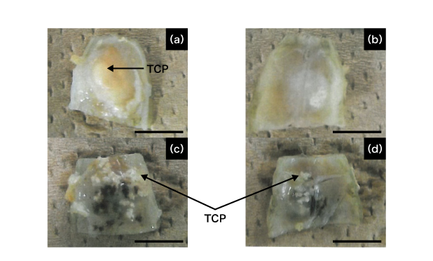
scale bar: 5.0mm
<a & b> "administration" group, <c & d> "non-administration" group
The pictures of the tissues around the bone defect parts of the "administration" group and the "non-administration" group are shown in Figure 2. The bone defect parts are seen between 2 lines in the pictures. Pink areas are bones, white areas are granular β-TCP, and light pink areas are fibrous or other tissues. In both groups, small new bones extending from the edge of existing bones to the center of the defect parts were found, and also remains of β-TCP granules were found because the β-TCP was not completely absorbed into the new bone. In the "non-administration" group, the new-bone formation was found at upper areas of the defect parts (areas in contact with the scalp) and at marginal areas of the defect parts (areas near the existing bones) only. However, in the "administration" group, it was also found at central areas of the defect parts in addition to above two areas.
Figure 2: Pictures of tissues at areas in the vicinity of defect parts of the calvaria in "administration" and "non-administration" group (x10)
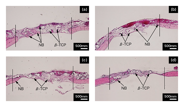
<a & b> "administration" group, <c & d> "non-administration" group,<NB> new bone
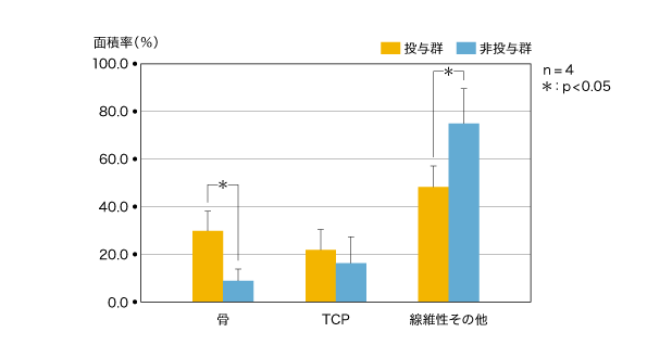
We took pictures of all the models for the whole image of the defect parts as shown in Figure 2, calculated the area ratio of the new bone, the β-TCP, and the fibrous or other tissues (n=4) , and then estimated them with statistical hypothesis at a significance level of 5%. The result is shown in Figure 3.
Figure 3: Comparison of area ratio of tissues in bone defect parts in "administration" and "non-administration" groups n=4, ∗: p < 0.05
3) Discussion

When CTP-containing collagen was administered orally to the rats implanted with granular β-TCP in the defect parts of their calvarias for 14 days, the osteogenesis at the areas in the vicinity of β-TCP were significantly hastened in comparison with the non-administration rats. It was a preferable outcome that the promotion of the osteogenesis was caused at the area away from the existing bones during the healing process of the bone. As a reason for the higher promotion of the osteogenesis around the bone prosthetic material (β-TCP), a possibility of synergism of 2 promoting actions was suggested; one that occurred in proliferation of the osteoblast brought by the β-TCP implant, and another that occurred in maturity of the osteoblast brought by the CTP-containing collagen administration.
