


Improvement of knee osteoarthritis (in vivo)
In a normal knee joint, the "articular cartilage" at the edge of the femur and the tibia works as a cushion, and its main component; type II collagen plays an important role. However, when a knee joint suffered osteoarthritis(OA), degradation of components of the articular cartilage caused by injury or aging that are associated with meniscus injury or ligament rupture leads to abrasion and collapse of the cartilage, and eventually it comes to cause outgrowth of bones around the joint (osteophyte formation) and disorder of joint functions (decreasing of sliding and cushioning functions) with accompanying synovitis. Then, we studied the effect of oral ingestion of CTP-containing collagen against destruction of the cartilage in OA.
Test for reduction of cartilage destruction in OA
1) Methods
OA model of male rabbits (at 13 weeks age) which were excised of their meniscus partially were prepared, and administrated test samples of 80 mg/kg per day for 5 weeks from 1 week before the model preparation day. Then, the areas around knee joints were removed 1 day after the final administration, and status of the articular cartilages was evaluated by histological observation. For the test samples, we prepared 2 types of CTP-containing collagen for verifying whether there was a difference in the effect depending on raw materials; a) CTP-containing collagen derived from pig skin collagen and b) CTP-containing collagen derived from fish scale. Moreover, we also prepared c) general collagen peptides derived from pig skin (CP) which did not include tripeptides for comparison. Adding a control group which was not administered any collagen peptides, the above 4 groups were examined.
2) Results

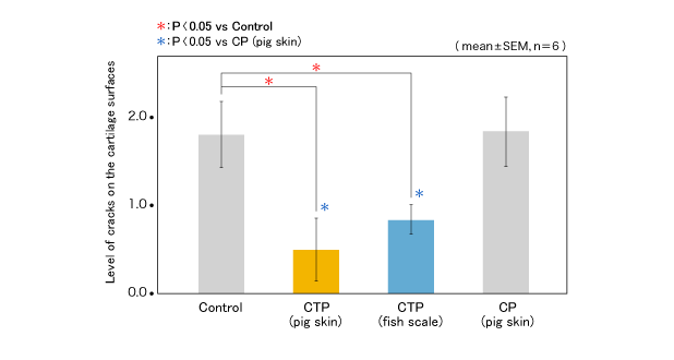
The tissue images of left tibias are shown in Figure 1. (Stained red parts are the cartilages.) Focusing on the outer articular cartilages which were the operation sites in model preparation, remarkable abrasion and elimination of the cartilage were observed in the control and c) CP groups, but remains of the cartilage were observed in the a) and b) CTP addition groups. In the inner articular cartilages on the opposite side, the cartilages were getting thin in the control and the c) CP groups, but a) and b) CTP groups showed quite thick cartilages (Figure 1). It was also confirmed that the level of cracks on the cartilage surface at the operation sites were significantly less in the a) and b) CTP groups than that in the control and the c) CP groups as a result of scoring and analysis of those cracks (Figure2).
The tissue images of left tibias (Triple staining by Safranin O, Fast green, and Iron -Hematoxlin)
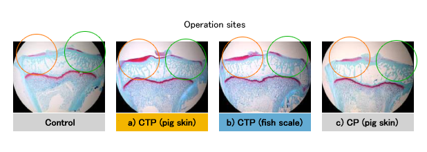
Figure 2: The levels of cracks on the cartilage surface (Tissue observation score)
3) Discussion

In the study with OA model rabbits, we verified the effect on reduction of cartilage destruction in OA by oral ingestion of CTP-containing collagen. Moreover, an apparent difference in the effect was observed in the presence of CTP, but no difference was observed based on the original raw material (pig skin/fish scale). Therefore, the study suggests that the bioactivity of CTP causes the effect on OA, regardless of the raw material from which the CTP is made.
